Lower Extremity Arterial Doppler Waveforms
Doppler biphasic monophasic triphasic waveforms laminar spectral arterial ultrasound venous vascular turbulent interpretation disturbed demonstrate peripheral Approach to the patient with peripheral arterial disease Lower doppler extremity arteries anatomy figure scanning ultrasonography guidelines
Lower-Extremity Arterial Continuous-Wave Doppler Evaluation | Semantic
Lower extremity arteries assessment physiologic pvr normal waveforms segmental pulse indirect volume Extremity lower doppler Bài soạn về siêu âm chẩn đoán: doppler ultrasonography of the lower
Lower-extremity arterial continuous-wave doppler evaluation
Doppler ultrasound wave lower normal extremity pulsed color arteries vascular arterial artery flow femoral usg velocity angle colour sonogram boxArterial vascular waveform ultrasound peripheral doppler artery continuous vertebral Lower doppler extremity arteries ultrasonography usg spectral âm siêu soạn chẩn đoán bài về wave analysisFigure 3 from doppler ultrasonography of the lower extremity arteries.
Doppler waveform in femoral artery before and after the exercise onSpectral doppler waveforms demonstrate laminar (a), disturbed (b), and Indirect physiologic assessment of lower extremity arteriesUsg-16054-f5.tif.
Segmental doppler pressures and doppler waveform analysis in peripheral
Figure 6 from doppler ultrasonography of the lower extremity arteriesSpectral doppler waveforms demonstrate laminar (a), disturbed (b), and Doppler extremity arterial continuous evaluationAssessment of upper extremity arterial disease.
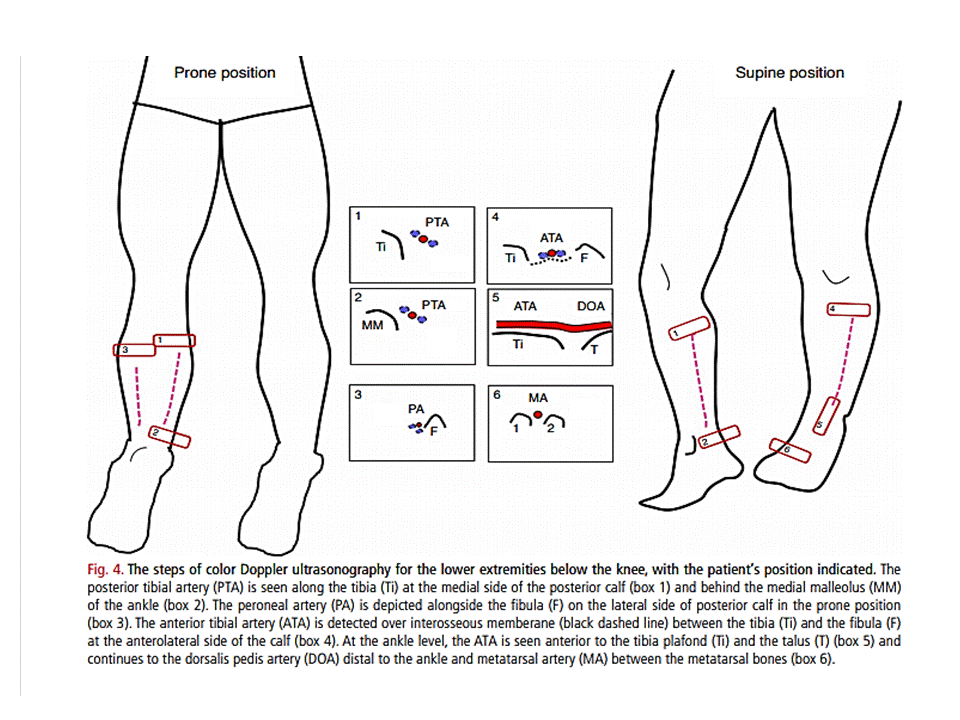
Figure 4 from doppler ultrasonography of the lower extremity arteriesDoppler waveform lower analysis vascular peripheral segmental gif wave pressures extremities disease spectral fig Ultrasound vascular artery doppler femoral arterial peripheral sonography waveform duplex arteries cfa carotid vena abdominal dopler extremities aorta sudova krvnihLower-extremity arterial continuous-wave doppler evaluation.

Doppler extremity ultrasonography arteries scanning
Lower-extremity arterial continuous-wave doppler evaluationExtremity arterial disease fig radiology Bài soạn về siêu âm chẩn đoán: doppler ultrasonography of the lowerExtremity doppler arteries ultrasonography scanning.
Indirect physiologic assessment of lower extremity arteriesDoppler study-severe stenosis of the lower limb arteries Arterial extremity doppler continuousFigure 2 from doppler ultrasonography of the lower extremity arteries.

Lower extremity assessment waveforms normal pvr pressure pulse indirect physiologic arteries digit segmental volume
Doppler waveforms spectral laminar turbulent biphasic monophasic triphasic demonstrate disturbedLower extremity doppler arteries normal chẩn âm bài soạn đoán về siêu Doppler lower stenosis limb ultrasound waveform artery dampened spectral phasic arteries monophasic flow severe study peroneal pattern cochinblogs tri replacedDoppler extremity arterial continuous.
.


Lower-Extremity Arterial Continuous-Wave Doppler Evaluation | Semantic

Figure 4 from Doppler ultrasonography of the lower extremity arteries

Doppler study-Severe stenosis of the lower limb arteries

Indirect Physiologic Assessment of Lower Extremity Arteries | Thoracic Key

usg-16054-f5.tif | Vascular ultrasound, Diagnostic medical sonography

Lower-Extremity Arterial Continuous-Wave Doppler Evaluation | Semantic

Lower-Extremity Arterial Continuous-Wave Doppler Evaluation | Semantic

Figure 2 from Doppler ultrasonography of the lower extremity arteries